X- Rays
Object and Mechanism Descriptions: An x-ray is a machine used for medical purposes in which it helps to see and detect injuries or any other irregularities found in the body. A concentrated beam of radiation is placed on any part of the body in which an image is produced where medics are able to see bones and dense tissues. There are a variety of x-rays in which some are specifically for dental purposes, arm x-rays, chest x-rays, etc. Most x-rays look like film projectors where one would see in class or watch a film. Just like a projector where there is a circular part which allows the light to go through and then projects it onto a screen… x-rays also have that part in which the concentrated beam is passed through.
The x-rays key characteristics of how it is built are: the motor, tungsten anode, vacuum, electron beam, oil bath, cathode, lead case, and the filter.
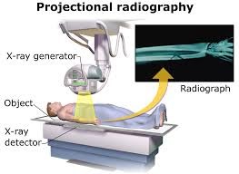
Process Descriptions: The main process of the x-ray is to help medics see internally for any abnormalities. The process of getting x-rays taken is when a patient starts to shows symptoms that they have not felt before. For example, if you start feeling chest pains making it hard for you to breath then it is important to get it checked out. The doctors will most likely send you to get a chest x-ray. The person who is getting the x-ray taken first has to be in a still position that they are told to be in so that the x-ray image is produced clearly. Then an apron mostly made of lead helps shield sensitive organs from the radiation, is placed on the patient. The authorized person who can operate the machine then clicks the button where the radiation is given only for a few seconds. Medics are then able to see the internal body by looking at the radiographic film.

This entry is licensed under a Creative Commons CC0 1.0 Universal Public Domain Dedication license.